
Diabetes is commonly linked to complications such as heart disease, stroke and kidney disease.
But its effects on the eyes can be just as serious.
Diabetic retinopathy develops when persistently high blood sugar levels damage the small blood vessels in the retina, the light-sensitive layer at the back of the eye.
The longer a person has diabetes, the higher the risk of diabetic retinopathy.
In its early stages, this condition may not affect vision or cause noticeable symptoms.
However, as it progresses, patients may experience floaters (dark spots or specks in their field of vision), blurred or distorted vision, and in severe cases, permanent vision loss.
Retired senior consultant ophthalmologist and former Health Deputy Director-General (Research and Technical Support) Datuk Dr Nor Fariza Ngah says a common misconception among people with diabetes is that clear vision means that their eyes are healthy.
Retinal damage, however, may already be present even when patients can still see clearly.
According to the 2023 National Health and Morbidity Survey (NHMS), an estimated 3.6 million people in Malaysia were living with diabetes, making regular eye examinations an important part of managing the condition.
“All diabetic patients will get diabetic retinopathy, but with different severity,” she says.
How does it happen?
Diabetes damages the retina through two processes: vascular leakage and vascular occlusion.
Dr Nor Fariza describes the former as affecting the veins and the latter involving the arteries.
Explaining vascular leakage, she says blood vessel walls normally act like pipes, allowing blood to flow without escaping into the surrounding tissue.
In people with diabetes, particularly those with uncontrolled blood sugar levels, pericytes – specialised cells that support small blood vessels – can become damaged and die.
“When this happens, fluid can leak out of the blood vessels into the surrounding tissue,” she says.
This leakage can be detected during retinal examinations, where doctors can identify fluid accumulation and deposits known as exudates.
In vascular occlusion, the blood vessels become narrow and might even become completely blocked.
When that happens, blood flow to certain areas of the retina is reduced, depriving the tissue of oxygen.
Dr Nor Fariza explains that the affected area first experiences hypoxia, or insufficient oxygen, before progressing to ischaemia, which occurs when blood supply is inadequate.
“When hypoxia occurs, the body releases certain substances to stimulate the formation of new blood vessels,” she says.
However, the newly-formed blood vessels do not function well and can contribute to bleeding inside the eye, she notes.
Why damage goes unnoticed
Diabetic retinopathy can go unnoticed because the damage does not necessarily affect the part of the retina responsible for central vision.
Dr Nor Fariza explains that this area, known as the macula, enables us to see objects clearly and sharply.
When damage or swelling affects the macula, patients may experience blurred or distorted vision.
If abnormalities occur elsewhere in the retina, their central vision may remain unaffected.
“They may still be able to see clearly and the condition may not cause any pain,” she says.
A retinal examination can reveal abnormalities such as bleeding and fatty deposits, even when patients have no visual complaints.
“That is why regular eye examinations are important.
“We cannot tell what is happening inside the eye simply by looking at it from the outside,” she says.
“By the time a diabetic patient comes to us with complaints such as blurred vision, red eyes or seeing what appears to be blood inside their vision, changes are likely to have already occurred in the eye,” she says.
In advanced cases, abnormal blood vessels may rupture, causing a vitreous haemorrhage, which can lead to sudden vision loss.
This can eventually lead to scar tissue formation, a process known as fibrosis.
Under the Health Ministry’s clinical practice guidelines, patients with diabetes are recommended to undergo retinal screening at least once a year.
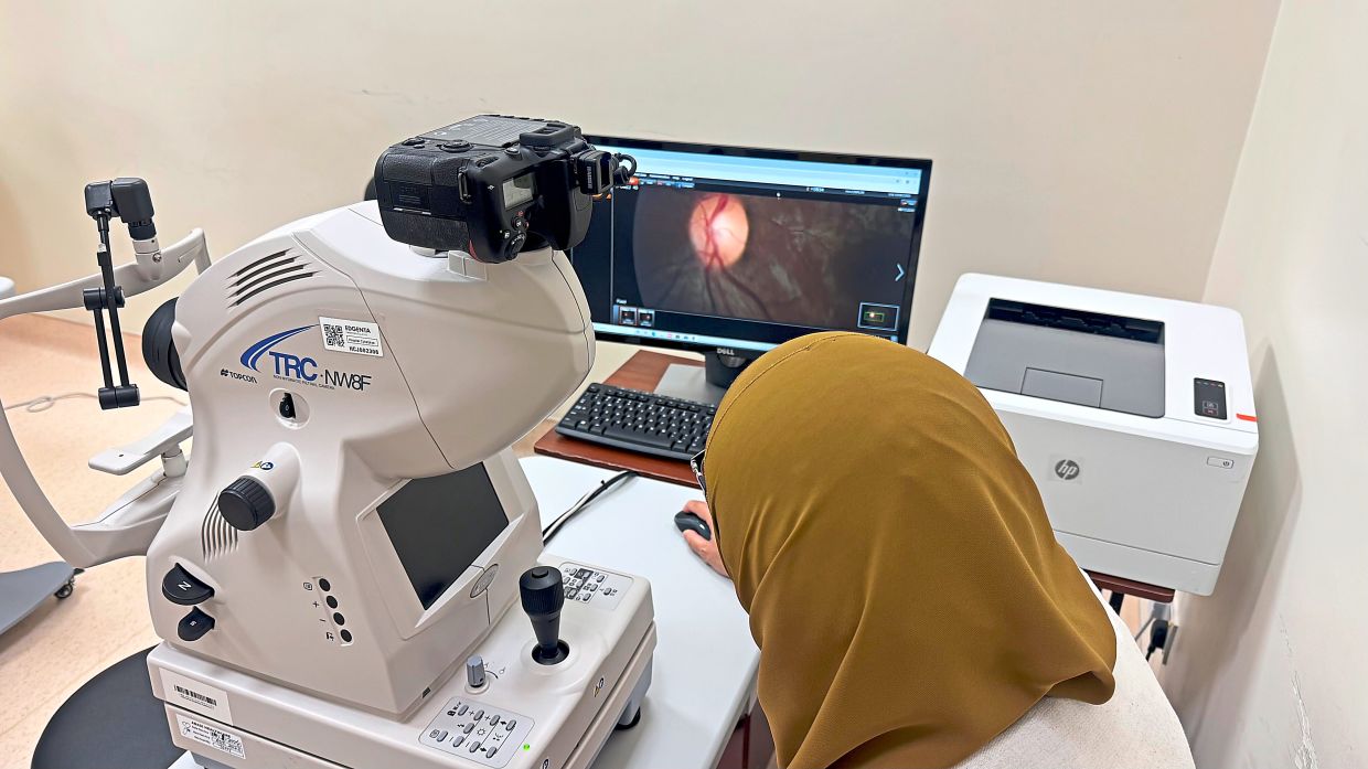
The screening uses a fundus camera to capture images of the retina, which are then examined for signs of diabetic retinopathy.
For those diagnosed with type 2 diabetes, Dr Nor Fariza advises having an eye examination at the time of diagnosis rather than waiting for symptoms to develop.
“Just because you have been diagnosed today does not mean the disease has only started today.
“You may have had diabetes for 10 years without knowing it,” she says.
AI speeds up screening
To improve retinal screening, the Health Ministry, in collaboration with technology partner Qmed Asia, developed an artificial intelligence (AI)-assisted system known as Diabetic Retinopathy Multi Artificial Technology Automation (Dr Mata).
The aim of the project is to support wider use of AI-assisted retinal screening in the public healthcare system.
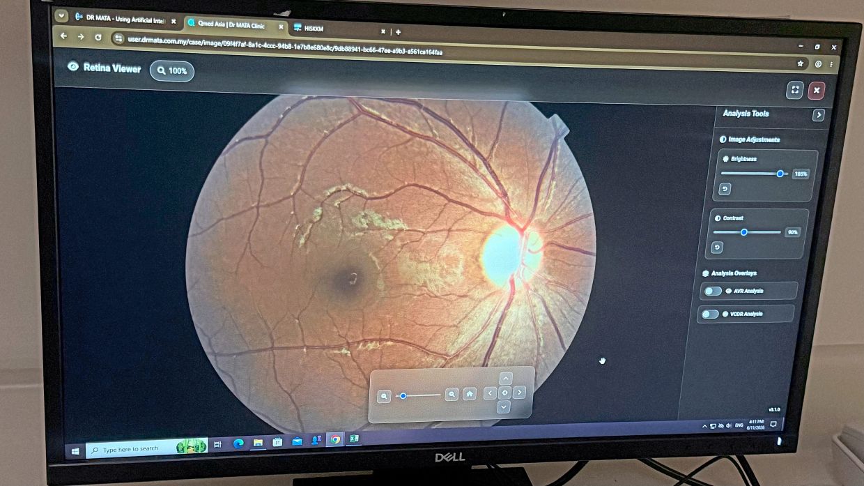
Senior consultant ophthalmologist and Hospital Cyberjaya Department of Ophthalmology head Dr Zalifa Zakiah explains that the system uses computers connected to fundus cameras to capture retinal images.
These images are then analysed by AI to identify abnormalities and grade the severity of diabetic retinopathy on a scale of 0 to 2.
The system can also automatically generate referral letters for patients who require further assessment by an ophthalmologist.
“Unlike systems built into specific camera models, Dr Mata operates as a web-based platform.
“Images captured using compatible fundus cameras are uploaded to the cloud for analysis, allowing the system to work with different camera brands,” Dr Zalifa explains.
Previously, retinal images taken at primary healthcare facilities had to be reviewed manually by trained healthcare personnel or specialists, Dr Nor Fariza explains.
This could lead to lengthy waiting times, particularly when the doctor trained to interpret the images was unavailable.
“Patients could wait between two weeks, and sometimes up to two months, to receive their results, delaying referrals for further assessment and treatment,” she says.
“With this system, we can take a picture and get the result in less than 30 seconds.”
During a one-month pilot programme in 2025, the system screened approximately 1,000 patients.
It initially recorded an accuracy rate of 83%, although Dr Nor Fariza says the AI’s performance has since improved.
Following the pilot project, selected primary healthcare facilities in Kedah and Perlis began using Dr Mata in January (2026).
The team hopes to expand the system to more healthcare facilities nationwide, particularly those already equipped with fundus cameras.
However, Dr Nor Fariza says wider implementation will depend partly on available funding, including resources for system maintenance.
Available treatments
Treatment for diabetic retinopathy depends on the extent of retinal damage and the areas affected.
According to Dr Nor Fariza, patients with milder disease may be monitored through regular examinations while managing their blood sugar levels and other risk factors, including high blood pressure and smoking.
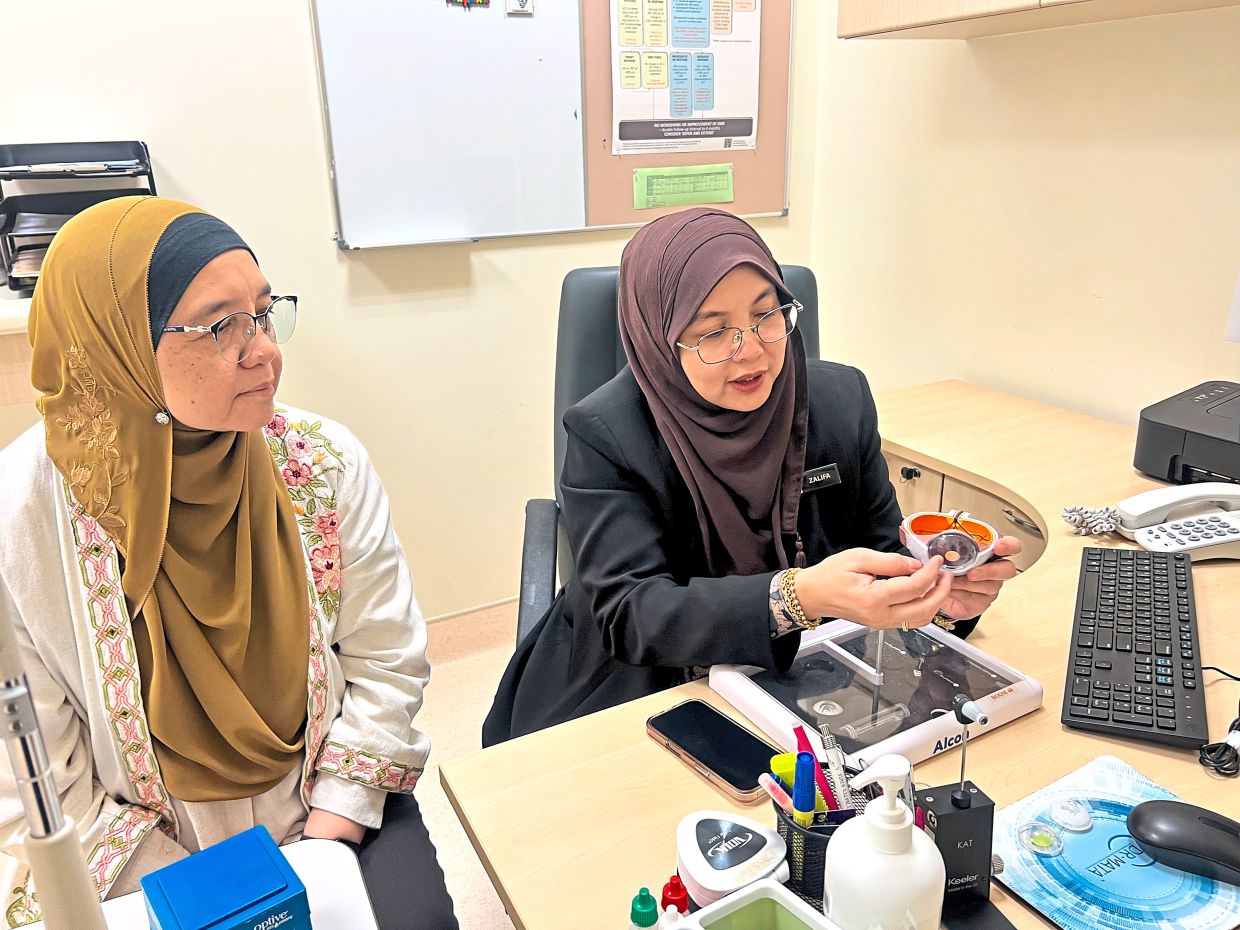
For more severe cases, panretinal photocoagulation (PRP), which is a laser treatment, has traditionally been one of the main methods of management.
When the retina is affected by ischaemia, laser treatment helps reduce its oxygen demand, suppressing signals that stimulate the growth of abnormal blood vessels.
For fluid leakage and swelling, Dr Nor Fariza explains that laser treatment is used to stimulate retinal cells, helping them absorb the accumulated fluid.
Another option for patients with diabetic macular oedema, where fluid accumulates in the macula and causes swelling, is an intravitreal anti-VEGF injection, which delivers medication directly into the eye.
She says that the medication helps reduce fluid accumulation and swelling.
In more advanced cases, where scar tissue develops and pulls on the retina, surgery may be necessary to remove the scar tissue.
This allows the retina to return to a more normal anatomical position.
However, restoring the retina’s structure does not necessarily mean its function or vision will fully recover.
“Sometimes, we can restore the anatomy, but in terms of function, we cannot be certain.
“If some of the retinal cells are already damaged or dead, we cannot bring them back and the patient may not regain their vision,” says Dr Nor Fariza.
Despite ongoing efforts to raise awareness, she says that many people with diabetes still underestimate the importance of eye health.
“They may think it is acceptable as long as they are not experiencing discomfort or pain,” she notes.
She adds that some patients are more afraid of dying or receiving a serious diagnosis, such as a heart attack or stroke, than losing their eyesight.
They may not realise how difficult life can become after losing their vision.
Unlike a patient who dies, someone who becomes blind continues to live, but may require lifelong care.
“It is not just the patient who is affected; the entire family is affected,” she points out.