

Robotic-assisted surgery is expanding treatment options for selected breast cancer patients, offering a minimally-invasive alternative to conventional surgery.
At Universiti Malaya Medical Centre (UMMC), the technology was introduced in 2024 for urological procedures before expanding to breast surgery last year (2025), according to consultant general and breast surgeon (oncoplastic) Assoc Prof Dr See Mee Hoong.
The system uses a specialised console and robotic instruments, providing surgeons with a magnified, three-dimensional (3D) view of the surgical area as they operate through small incisions.
Although the technology can enhance precision and instrument control, it does not operate independently or make surgical decisions.
“The surgeon remains responsible for everything, including decisions about the patient’s surgery and the outcome.
“The robot simply helps to enhance the surgery,” Assoc Prof See says.
What it’s used for
At UMMC, robotic-assisted breast surgery is used mainly for nipple-sparing mastectomy.
This procedure removes breast tissue while preserving the skin and nipple-areolar complex, provided it is oncologically safe (meaning that it does not compromise cancer control or increase the risk of recurrence).
For this reason, Assoc Prof See says careful patient selection is essential.
She explains that the tumour should generally be more than 3mm to 5mm from the skin and should not invade the underlying muscle.
Selected patients with T1 tumours (up to 2cm) or T2 tumours (between 2cm to 5cm) may also be suitable.
The technique may be an option for patients who are unsuitable for breast-conserving surgery, including those with extensive areas of microcalcifications – tiny calcium deposits in the breast that can sometimes indicate early breast cancer – even in stage 0 disease.
However, patients who require more extensive surgery may not be suitable for this minimally-invasive approach.
Assoc Prof See says the decision depends on three main factors: patient-related factors, the characteristics of the cancer, and personal preferences for breast reconstruction.
“The objective is very crucial here. It’s for effective cancer treatment,” she says, adding that the aim is also to improve patients’ appearance and quality of life.
Nipple-sparing mastectomy is often followed by immediate breast reconstruction using an implant.
However, some patients choose not to undergo reconstruction – an approach commonly known as “going flat”, she says.
“We preserve the skin and nipple-areolar complex, but there is no additional reconstruction or anything inserted underneath the skin.”
For women whose breasts have little projection, the difference in appearance without reconstruction may be less noticeable, she says.
Robotic-assisted nipple-sparing mastectomy may also be considered for selected individuals with inherited BRCA1 or BRCA2 gene mutations who choose to have their breast tissue removed to reduce their risk of developing breast cancer before receiving a diagnosis.
Reconstruction may follow, using an implant or the patient’s own tissue.
Evolution of breast cancer surgery
Breast cancer surgery has evolved towards less invasive techniques that aim to preserve patients’ appearance without compromising cancer treatment.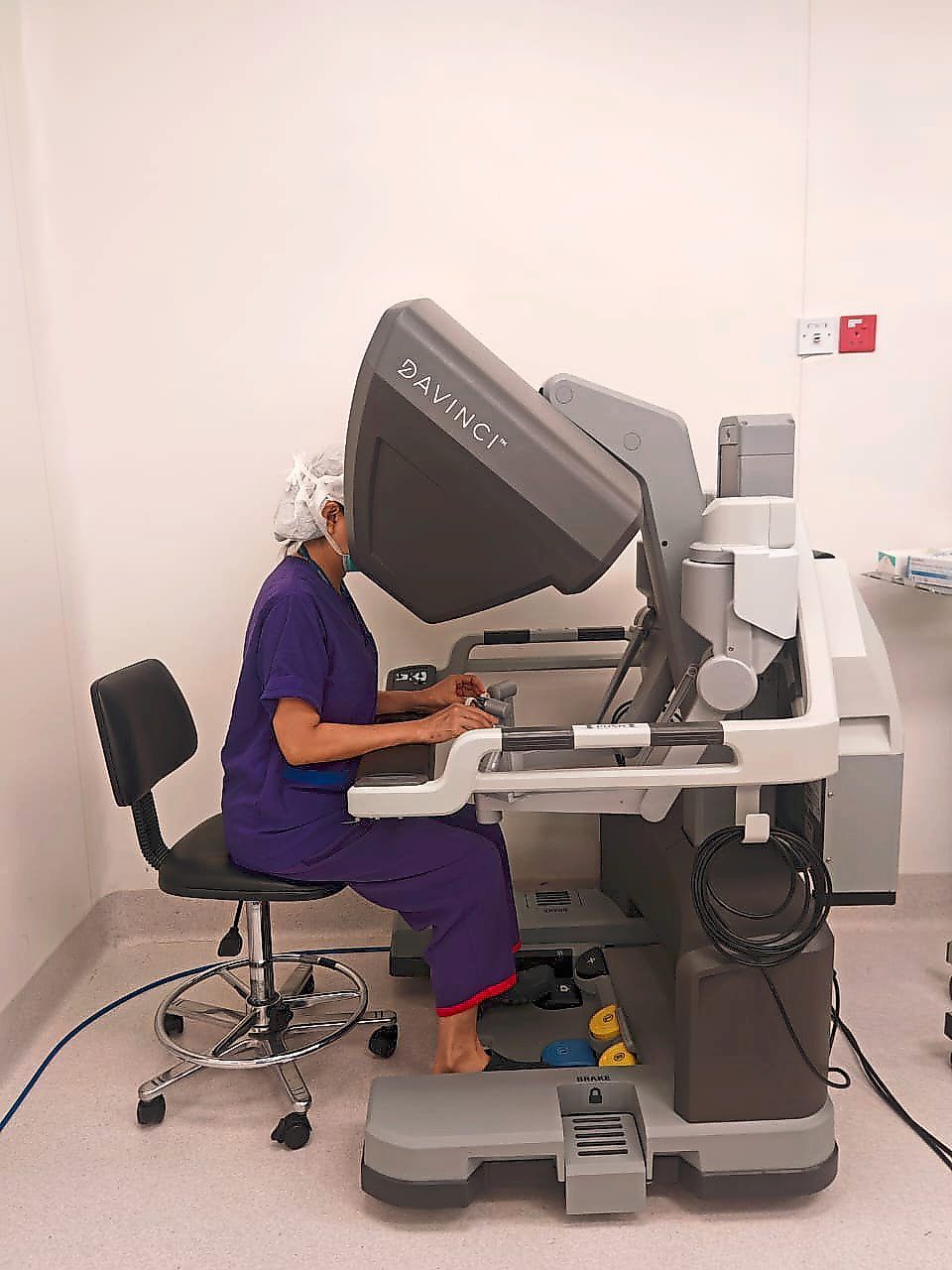
According to Assoc Prof See, conventional open mastectomy typically leaves a scar across the chest, extending from near the midline towards the side.
“The scar can be more than 10cm,” she notes.
In the past, surgeons performed radical mastectomy, which involved removing the entire breast, nipple-areolar complex, underlying chest muscles and lymph nodes under the arm.
“At that time, you had to remove the muscle and everything, which could lead to serious shoulder complications,” she says.
Less extensive operations later became more widely adopted.
Modified radical mastectomy preserves the chest muscles while removing the breast and underarm lymph nodes, reducing the extent of surgery compared with radical mastectomy.
Assoc Prof See says that despite these advances, conventional surgery can still leave prominent scars and cause postoperative pain and changes in appearance.
“Some also develop hypertrophic scars or keloids, which can be itchy and painful,” she says.
To reduce visible scarring, surgeons have adopted minimally-invasive techniques that use smaller incisions, often at the side of the chest or below the armpit.
Endoscopic instruments allow breast tissue to be removed through these incisions while preserving the skin, and in suitable cases, the nipple-areolar complex.
Robotic-assisted surgery builds on this approach, using a magnified 3D view and articulated instruments that allow surgeons to manoeuvre with greater control.
“Even if the surgeon has some tremor or shaking of the hand, the robotic system can reduce that tremor,” says Assoc Prof See.
She says that the approach can help achieve favourable cosmetic outcomes, including less conspicuous scars and preservation of breast shape.
However, robotic assistance does not eliminate surgical risks.
She points out that complications can include bleeding, infection and damage to the blood supply of the nipple-areolar complex or surrounding skin, potentially leading to tissue necrosis, in which the affected tissue dies due to inadequate blood supply.
The surgical approach also involves a trade-off in operating time.
“Conventional surgery may take about an hour, whereas nipple-sparing mastectomy using this technique may take two to three hours because surgeons have to work carefully through smaller incisions,” she says.
Recovery, however, varies depending on the individual patient and the extent of surgery.
Assoc Prof See says smaller incisions may mean less post-operative pain and easier wound care.
“If the wound is smaller, patients generally experience less pain after surgery, and wound care is easier to handle,” she explains.
At UMMC, patients typically stay in the hospital for two to five days if there are no complications.
Assoc Prof See says minimally-invasive approaches may also help ease pressure on hospital beds by allowing suitable patients to recover at home, leaving beds available for those who need more intensive care.
Helping lower income patients
For lower-income patients, the cost of breast cancer tests and treatment can become a huge barrier to timely care.
Established at UMMC in 2017 and led by Assoc Prof See, the B40 Diagnostic Fund helps eligible patients access investigations such as mammograms, ultrasound scans and biopsies, which they may not otherwise be able to afford.
Some patients require more specialised procedures, such as MRI-guided biopsies, which can cost more than RM3,000, she says.
“Some of our patients have subtle changes that can only be detected through MRI [magnetic resonance imaging], which is why the scan remains important,” she notes.
Stereotactic mammography-guided vacuum-assisted biopsies, which use X-ray imaging to pinpoint suspicious breast abnormalities and collect tissue samples for testing, can also cost thousands of ringgit.
To date, the B40 Diagnostic Fund has helped more than 500 UMMC patients since it was first established.
UMMC has also introduced the B40 Robotic Surgery Fund to help eligible patients access robotic-assisted breast surgery.
Assoc Prof See says the initiative received a RM250,000 donation from Yayasan Canone Kianjoo to kickstart the funding.
“Robotic-assisted breast surgery costs about RM22,000 or more, excluding subsequent treatment, which makes it a significant expense for lower- income patients.
“Overcoming this financial barrier is not easy in our setting, and we hope to establish partnerships with more corporate organisations over time,” she says.
The hospital is also exploring ways to secure medical insurance coverage for the procedure.
Annual fundraising helps sustain the fund, including through the recent Pink ESG Wellness Campaign held in October (2026).
Assoc Prof See hopes that both funds will help more patients overcome financial barriers to treatment.
“The true measure of medical progress is not how advanced our technology becomes, but how many lives we can meaningfully improve.
“No woman should be denied appropriate breast cancer care simply because she cannot afford it,” she says.
She shares that lower-income patients who wish to access the B40 Diagnostic Fund must first undergo UMMC’s standard clinical assessment to determine the investigations they need.
They must also be referred to the hospital’s social welfare department to verify their B40 status.
Assoc Prof See says approval can be granted within one to two hours, although the process may take up to two days in more complex cases.
“This is beneficial for both patients and clinicians because financial concerns can cause patients to go home and delay further investigations, making it difficult for us to follow up with them promptly,” she says.
Those interested in supporting the Funds can email ppumcare@ummc.edu.my.